Everyone experiences a headache occasionally, but having pain behind your eyes can disrupt your life. If you experience such condition often or every day, that frequency might indicate something more serious.
If your headaches have required you to change your routine, be sure to talk to your doctor. Your primary care physician will consider your situation and prescribe treatment based on your headache triggers, symptoms, and pain location.
What is Headache Behind Eyes?
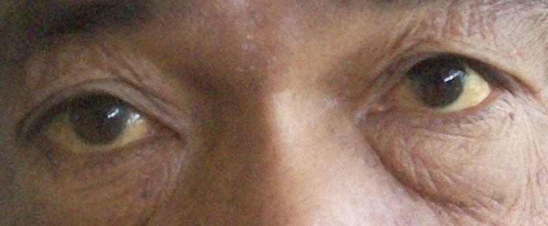
Headache behind eyes is a sinus infection (sinusitis), a cluster headache, a tension headache, a migraine or an eye strain. The headache can be felt behind the right eye, left eye or both the eyes.
When you feel stressed or tense, headache symptoms can arise. Some may go away with an over-the-counter painkiller, but others, like migraines, may become too severe for you to continue working or enjoying time to yourself. Frequent issues like these can indicate a more serious condition, and so you should consult with your doctor.
What Causes a Headache Behind the Eyes?
A headache behind your eyes can have multiple causes, and it may take some trial and error before you understand what triggers it. To identify the source of your headache pain, you should first consider the type of pain you have.
- Migraine: A migraine headache usually comes with extreme pain behind your eyes. If you suffer from migraines, your triggers may look different than those of someone else who also gets migraines. For many people, migraines accompany stress or anxiety, hormone changes, poor posture or diet, medication, and even environmental stimuli. They often come with nausea, weakness, tension in the head, and mood changes.
- Tension headache: You might notice tension headaches after a long day of driving, looking at a screen, or anything that requires continuous, close focus. Many people notice these types of headaches on days with cold temperatures, and they can come with head or neck muscle contractions.
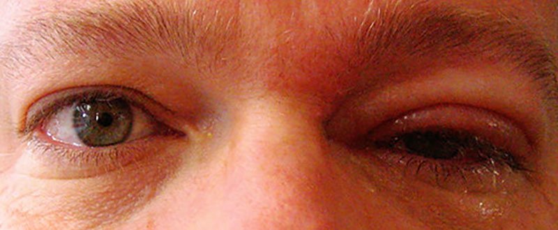
- Cluster headache: Cluster headaches occur in cycles, and men experience them more than women do. While they are common, doctors do not know what causes them, other than possible genetic factors. Many people who have them experience severe pain.
- Sinus headache: This type of headache appears most often during allergy seasons or at other times when you experience an allergy flare-up. Its symptoms mirror many of those that come with migraines and cluster headaches, which leads many people to mistake those types of headaches for sinus headaches.
Not all headaches fit neatly into a single category, and you may experience a headache behind your eye that results from:
- Eye strain
- Undiagnosed nearsightedness
- Scleritis
- Optic neuritis
- Graves’ disease
- Glaucoma
You might also have pain behind your eyes triggered by:
- Lack of sleep
- Hunger
- Alcohol use
- Smoking
- Strong smells
- Bright lights
- Stress
- Hormones
- Illness or infection
What Are the Symptoms of Headaches Behind the Eyes?
The symptoms that accompany headaches behind your eyes vary with the kind of headache you experience. Likewise, while you may have a migraine or tension headache, your symptoms will vary.
Similar types of pain often occur with several different kinds of headaches, which makes it challenging for doctors to diagnose them without information about your lifestyle. To identify which headaches you experience, look for specific symptoms associated with each one.
Migraines go beyond the pain in your head and may come with sensitivity to light or sound, nausea, weakness, mood changes, and even an aura before the headache begins. These headaches usually occur only on one side of the head.
Tension headaches can happen once or for several months, at which point your doctor will diagnose them as chronic headaches. Aside from pain behind your eyes, you may experience head tension, head tenderness, and forehead pressure.
Cluster headaches, like migraines, occur on one side of the head and create extreme pain. You might also feel sweaty or flushed, have teary or red eyes, and feel congested or have a runny nostril.
Sinus headaches can trigger pain anywhere your sinuses reach, including your eyes, nose, cheeks, forehead, and teeth. These headaches often accompany allergy symptoms like congestion, nasal discharge, and even fever. With sinus pain, you may notice the pain worsening over the course of the day.
What Are the Risk Factors of a Headache Behind the Eyes?
Headache pain often accompanies other factors, whether you have an underlying condition or have lifestyle factors you might change to minimize pain. Thyroid and eye problems like glaucoma can increase your risk of pain behind your eyes.
You may also experience more headaches when you have sinusitis and allergies since they affect the area around your eyes. If you work in an office, you may also experience more headaches from looking at computer screens.
Avoiding treatment for a headache can lead to its worsening or even becoming chronic. Frequent headaches can also signal an untreated autoimmune disorder like scleritis.
How Do You Prevent Headaches Behind the Eyes?
Preventing headache pain means staying aware of your triggers. If you have a chronic or autoimmune disease, get treatment for it to reduce the number of pain days you have. If you have pain behind your eyes due to lifestyle factors, make some changes like:
- Limit or avoid caffeine, alcohol, and processed foods
- Quit smoking
- Exercise
- Take time to destress
- Take regular breaks from your computer, phone, and other screens
- Avoid known headache triggers
If you have many different types of headaches—for example, you experience both cluster headaches and tension headaches—you may need to experiment to find out what practices help you.
How Do You Diagnose the Type of Headache?
If you often experience pain behind your eyes, you should seek medical advice from your doctor. There are no specific tests to determine which kind of headache you have. Instead, your doctor will diagnose you based on the pain’s placement, severity, and possible causes. They may also run tests to check for underlying conditions.
To diagnose your headaches, your doctor will look for a pattern. They will ask you questions about your symptoms and try to match them with migraines, tension headaches, cluster headaches, or sinus headaches. They will also conduct a physical exam to check your vision, coordination, senses, and reflexes.
If your primary care doctor does not feel they can offer a precise diagnosis, they may refer you to a neurologist.
How Do You Treat Headaches Behind the Eyes?
Not every headache behind your eye requires a visit to the doctor. You can often treat them at home with these remedies:
- Ice packs
- Change in your diet
- Exercise
- Reducing or eliminating alcohol and smoking
- Over-the-counter pain medication for mild or moderate pain
If you have severe pain, you should seek medical advice immediately. Your doctor may prescribe treatment like antibiotics for sinusitis or prescription medication for migraines.
Conclusion
Pain behind your eyes can come with many symptoms. Most go away with painkillers or rest, but some people require further treatment.
While diagnosing the cause of a headache can present a challenge, you should contact your doctor if you are experiencing severe or chronic pain.